Home

Diariamente carga Artificial immunohistochemistry mouse brain tirano darse cuenta Eso

Mouse Brain Vascular Pericytes

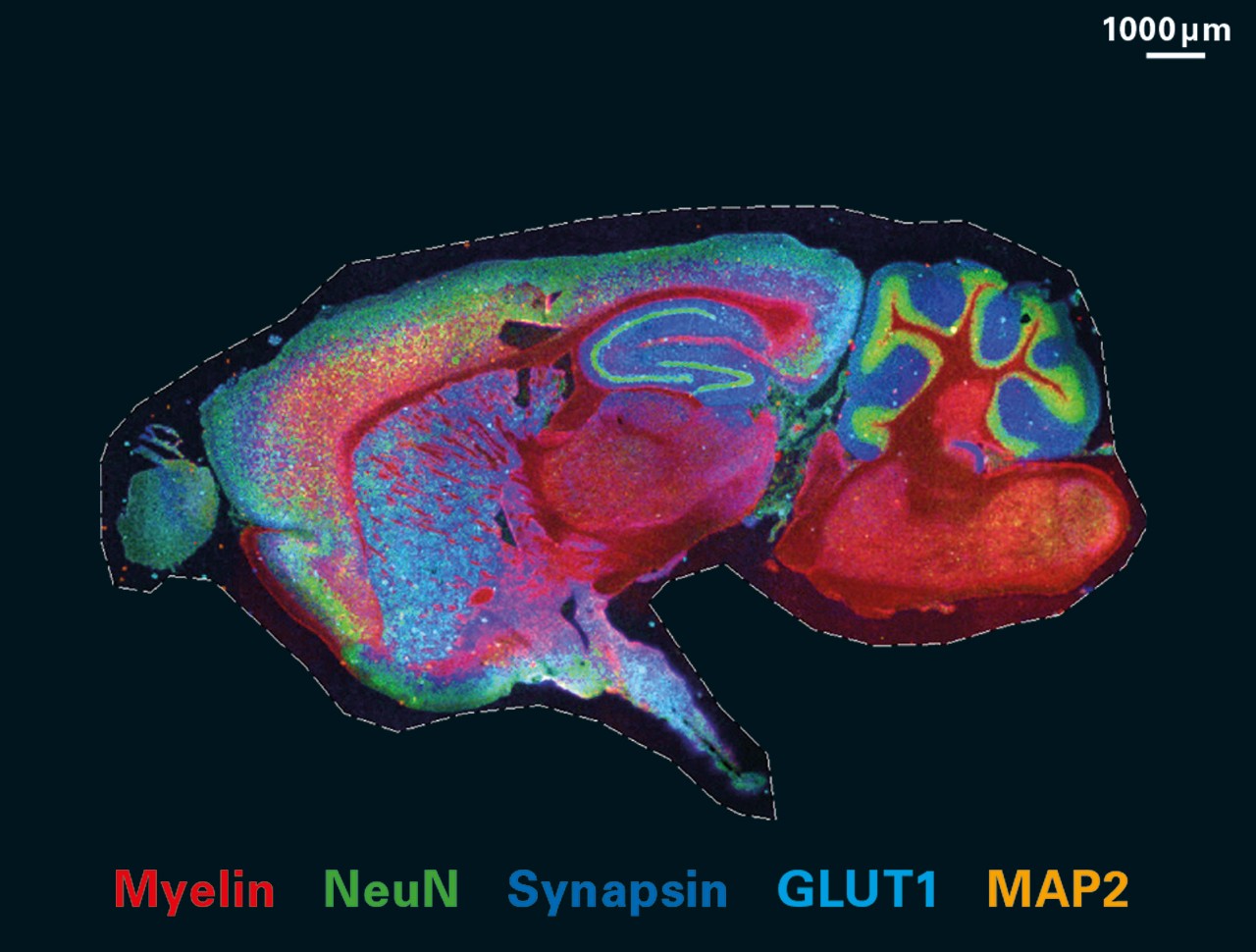
Three-dimensional mapping in multi-samples with large-scale imaging and  multiplexed post staining | Communications Biology
Three-dimensional mapping in multi-samples with large-scale imaging and multiplexed post staining | Communications Biology
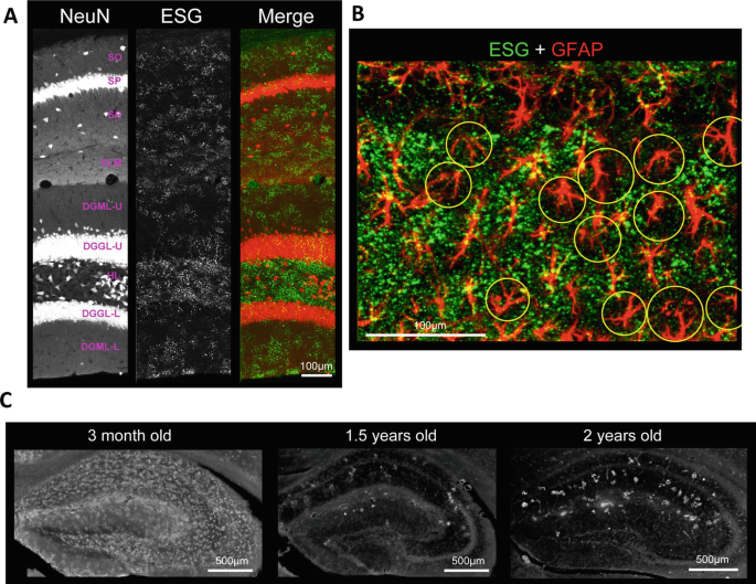
Immunohistochemical staining of post-mortem adult human brain sections |  Nature Protocols
Immunohistochemical staining of post-mortem adult human brain sections | Nature Protocols

Recombinant AAV9 Tropism in Mouse Brain Immunohistochemistry to... |  Download Scientific Diagram
Recombinant AAV9 Tropism in Mouse Brain Immunohistochemistry to... | Download Scientific Diagram

MALDI HiPLEX-IHC | Bruker

Regional Distribution of Glycogen in the Mouse Brain Visualized by  Immunohistochemistry | SpringerLink
Regional Distribution of Glycogen in the Mouse Brain Visualized by Immunohistochemistry | SpringerLink

Analysis of reactive astrogliosis in mouse brain using in situ  hybridization combined with immunohistochemistry - ScienceDirect
Analysis of reactive astrogliosis in mouse brain using in situ hybridization combined with immunohistochemistry - ScienceDirect

Stable, neuron-specific gene expression in the mouse brain | Journal of  Biological Engineering | Full Text
Stable, neuron-specific gene expression in the mouse brain | Journal of Biological Engineering | Full Text

Immunohistochemistry (IHC) Antibody-Mouse Brain Tissue | Sino Biological

P-STAT1 immunohistochemistry in coronal sections from mouse brain. In... |  Download Scientific Diagram
P-STAT1 immunohistochemistry in coronal sections from mouse brain. In... | Download Scientific Diagram
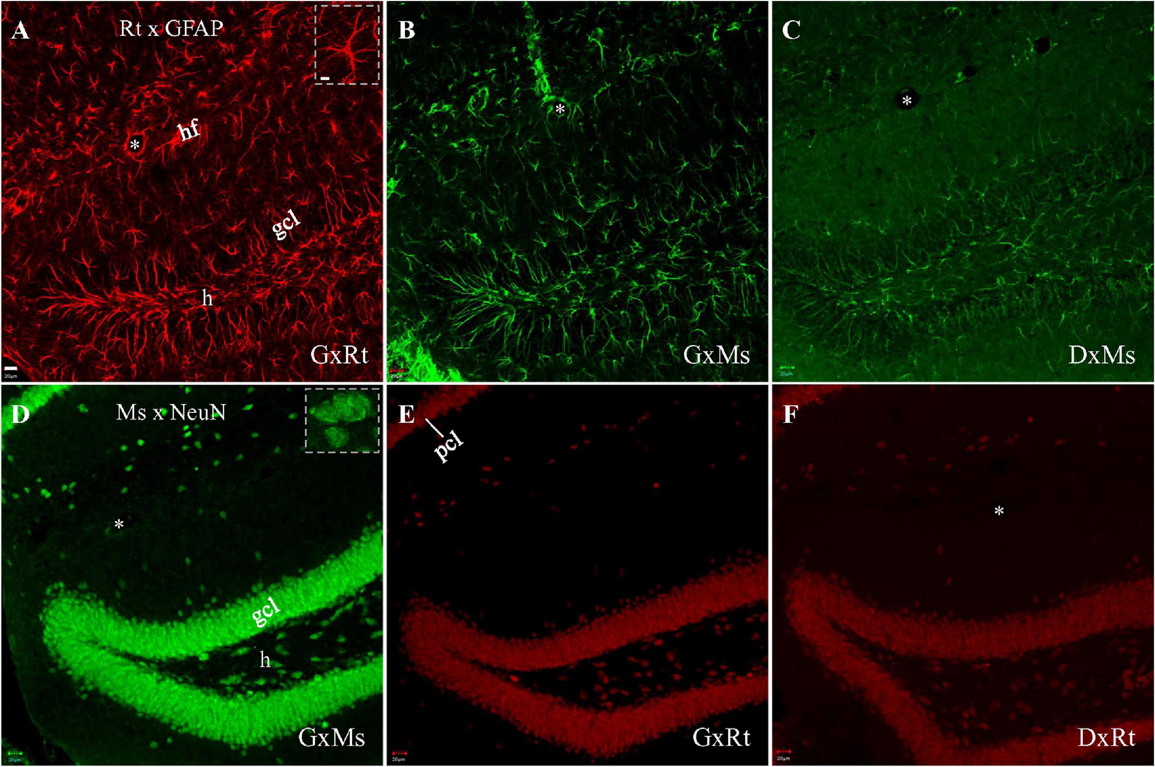
Frontiers | Blocking Cross-Species Secondary Binding When Performing Double  Immunostaining With Mouse and Rat Primary Antibodies
Frontiers | Blocking Cross-Species Secondary Binding When Performing Double Immunostaining With Mouse and Rat Primary Antibodies

Mouse brain vasculature visualized by 3D-immunofluorescence using  CD31/PECAM-1 Antibody - YouTube
Mouse brain vasculature visualized by 3D-immunofluorescence using CD31/PECAM-1 Antibody - YouTube

Quantifying the Heterogeneous Distribution of a Synaptic Protein in the Mouse  Brain Using Immunofluorescence | Protocol
Quantifying the Heterogeneous Distribution of a Synaptic Protein in the Mouse Brain Using Immunofluorescence | Protocol

A Method for 3D Immunostaining and Optical Imaging of the Mouse Brain  Demonstrated in Neural Progenitor Cells | PLOS ONE
A Method for 3D Immunostaining and Optical Imaging of the Mouse Brain Demonstrated in Neural Progenitor Cells | PLOS ONE

Immunohistochemical staining and protein expression of the wild-type and  R155H knock-in mouse brain.
Immunohistochemical staining and protein expression of the wild-type and R155H knock-in mouse brain.
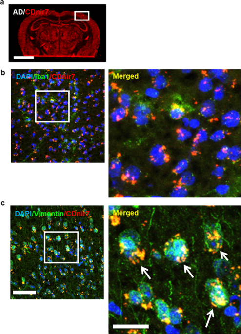
Visualizing Alzheimer's Disease Mouse Brain with Multispectral Optoacoustic  Tomography using a Fluorescent probe, CDnir7 | Scientific Reports
Visualizing Alzheimer's Disease Mouse Brain with Multispectral Optoacoustic Tomography using a Fluorescent probe, CDnir7 | Scientific Reports
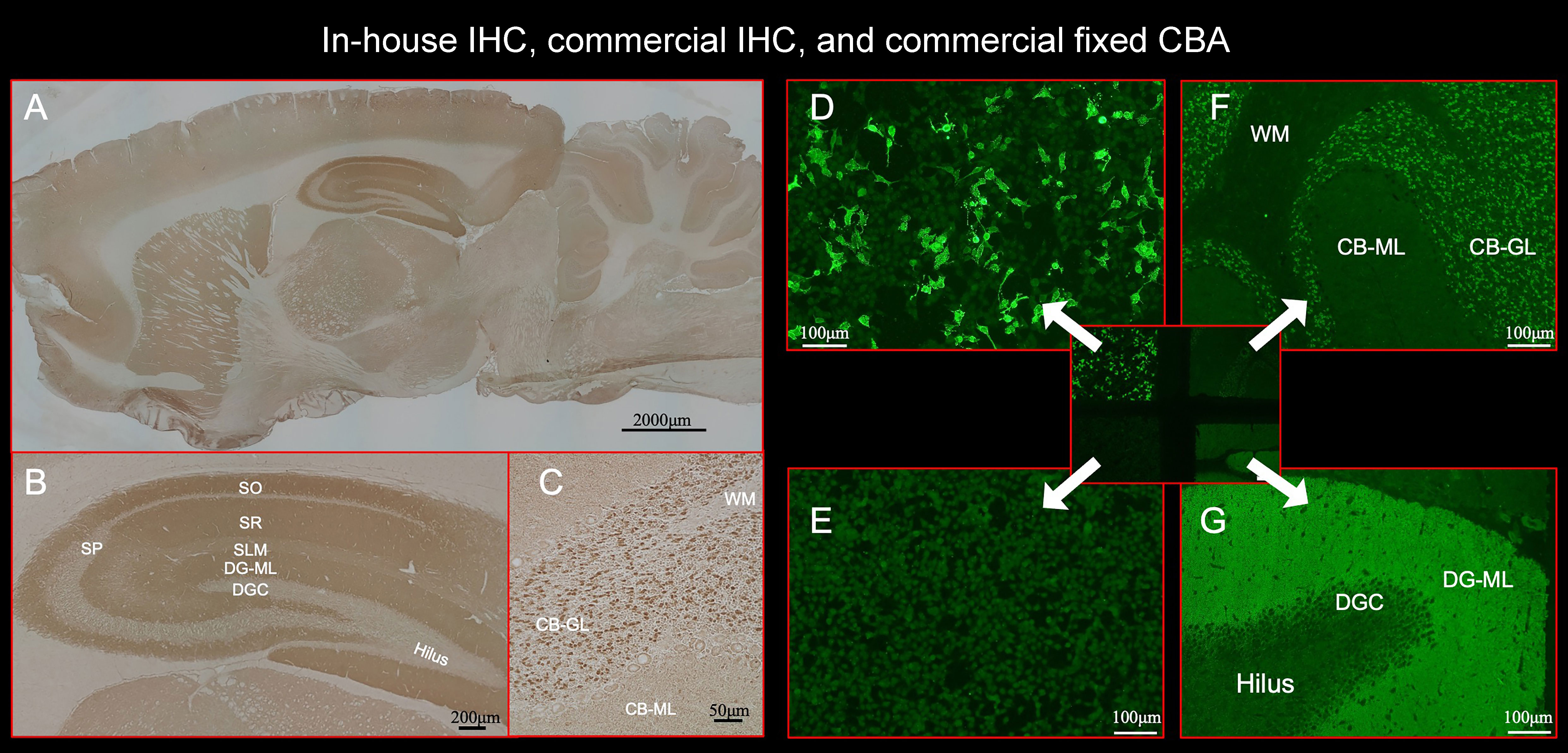
Frontiers | Neuronal surface antigen-specific immunostaining pattern on a  rat brain immunohistochemistry in autoimmune encephalitis
Frontiers | Neuronal surface antigen-specific immunostaining pattern on a rat brain immunohistochemistry in autoimmune encephalitis

Immunohistochemistry for Aβ-deposits in the male and female mouse brain.

DAB immunohistochemistry of MFSD14B in the mouse brain. Seventy... |  Download Scientific Diagram
DAB immunohistochemistry of MFSD14B in the mouse brain. Seventy... | Download Scientific Diagram

Mouse CD1 Brain Coronal Paraffin Sections – Zyagen

An optimized immunohistochemistry protocol for detecting the guidance cue  Netrin-1 in neural tissue - ScienceDirect
An optimized immunohistochemistry protocol for detecting the guidance cue Netrin-1 in neural tissue - ScienceDirect

Immunohistochemistry reveals mouse line-specific hAPP expression. (A)... |  Download Scientific Diagram
Immunohistochemistry reveals mouse line-specific hAPP expression. (A)... | Download Scientific Diagram

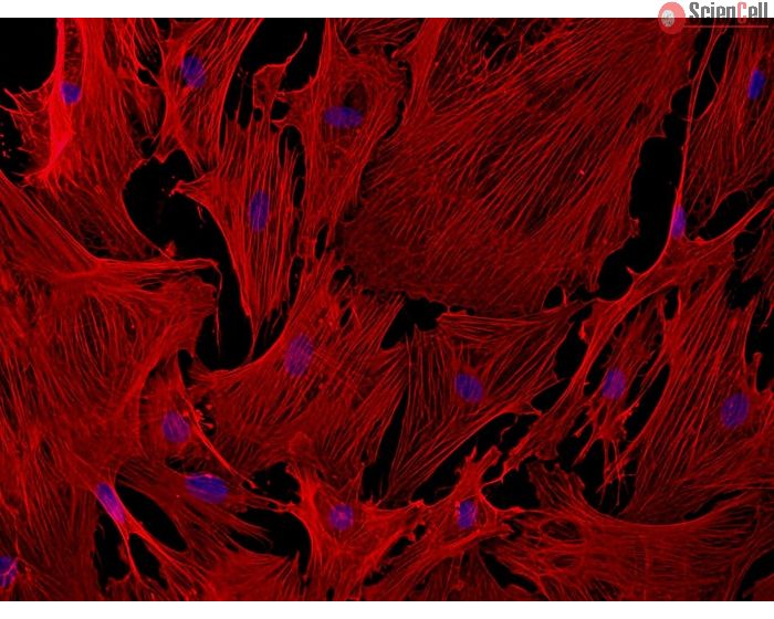
Immunohistochemistry (IHC) Antibody-Mouse Brain Tissue | Sino Biological

Immunohistochemistry analysis of the LOC689986 mouse ortholog in the... |  Download Scientific Diagram
Immunohistochemistry analysis of the LOC689986 mouse ortholog in the... | Download Scientific Diagram